Royal Hues Beneath the Lens
From Sacred Dyes to Cellular Stains

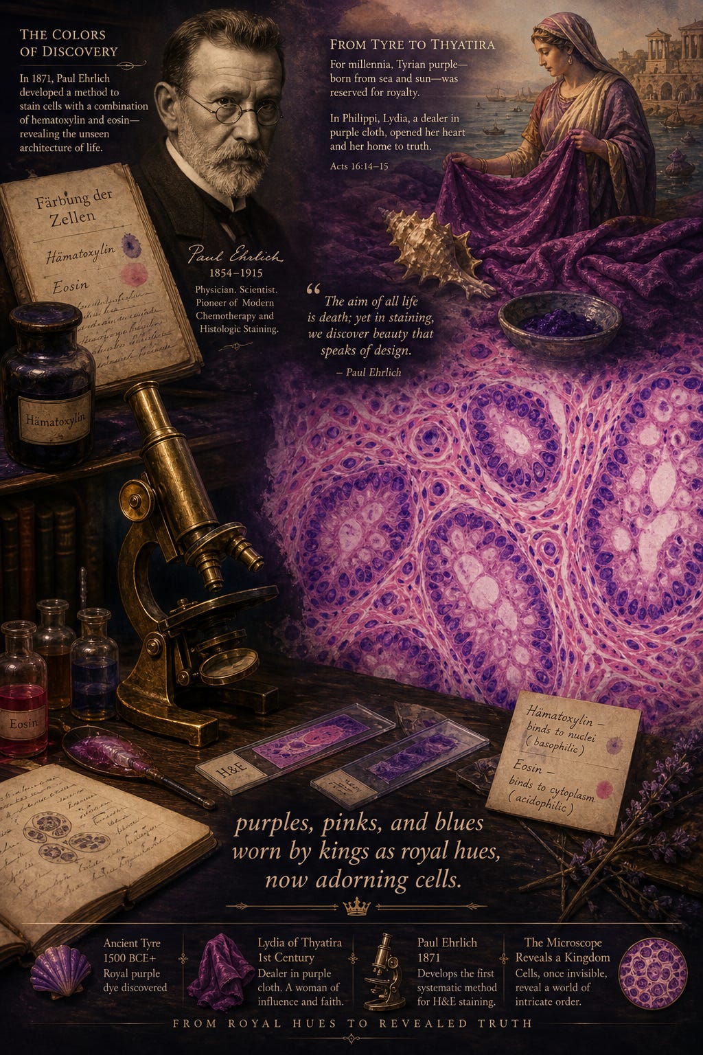
“purples, pinks, and blues
worn by kings as royal hues,
now adorning cells.”
I wrote this haiku years ago while a professor teaching medical students, and it was later published in the UCSD School of Medicine magazine.
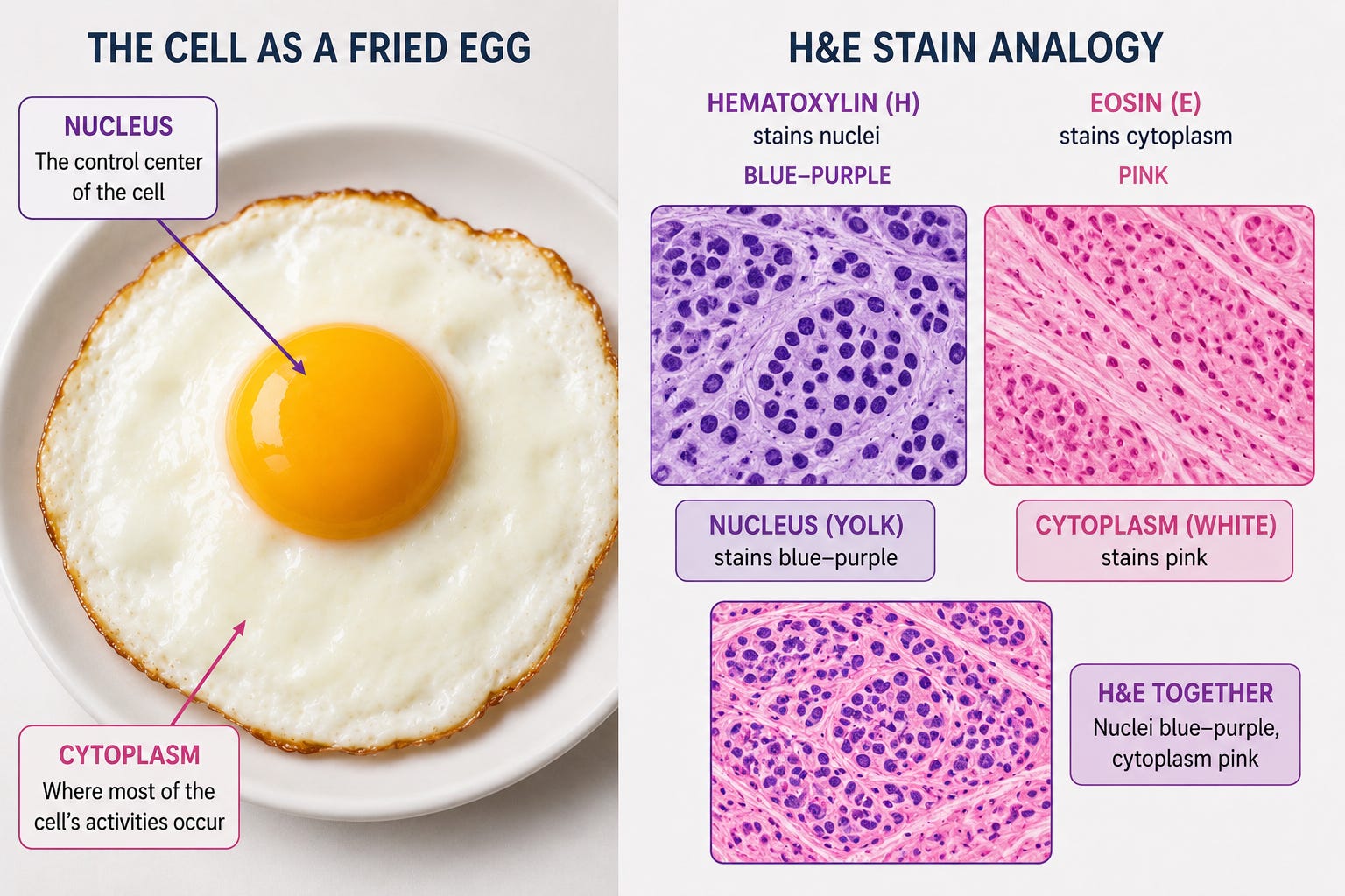
Hematoxylin and eosin — the classic H&E stain used in pathology — transformed medicine by allowing us to see the architecture of life through color.
Hematoxylin stains nuclei deep blue-purple, drawing attention to the genetic and structural center of the cell. Eosin stains the surrounding cytoplasm, connective tissue, muscle, and proteins in shades of pink, rose, and coral.
One of the earliest ways many medical students learn this is through the image of a fried egg. The nucleus is the yolk and the cytoplasm the surrounding egg white.
Under the microscope colors become a sophisticated visual language.
Pathologists recognize subtle shifts in hue, texture, or contrast, to distinguish normal from atypical which could be due to reactive, inflamed, premalignant, or worse yet malignant tissue.
Many of these staining techniques emerged during the great explosion of microscopy and cellular pathology in the late nineteenth century. Scientists and physicians experimented with textile dyes, coal tar derivatives, and plant pigments to add color to different parts of cells and tissues.
Paul Ehrlich, a German physician, scientist, and researcher, was one of the pioneering figures that helped develop staining methods that transformed microbiology, hematology, and histopathology in the late 1800s.
His work showed that different cellular structures have chemical affinities for dyes, allowing disease itself to become visually recognizable.
Later stains such as Wright-Giemsa and Diff-Quik expanded this visual language even further, especially in hematology and cytology. A drop of blood, a lymph node aspirate, or a thyroid specimen could now reveal inflammatory cells, parasites, bacteria, lymphoma, or metastatic cancer through subtle shifts in color and morphology.
Over time, the slide stops looking like “just tissue.” It begins to resemble landscapes, flowing rivers, woven fabric, stained glass, or galaxies unfolding in purples, pinks, blues, and crimson.
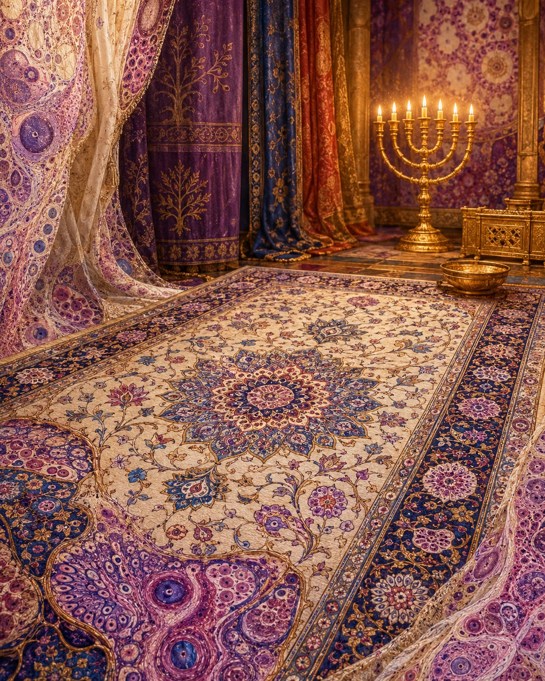
The Woven World
Persian carpets carry one of the oldest visual languages in the world. The earliest surviving pile carpet is more than two thousand years old, and by the Safavid period Persian weaving had become one of the great courtly arts, filling wool and silk with gardens, animals, vines, borders, medallions, and images of paradise.
Naeen’s own rug tradition appears to be more recent, emerging in the early twentieth century, but that makes my mother’s memory even more striking. She told me my grandmother had helped bring carpet making there. So the story is not only about ancient Persian art. It is also about a woman, a family, a region, and the patient transmission of beauty by hand.
Before I ever looked through a microscope, I learned to enter color through Persian rugs.
My grandmother made a small prayer rug. As a child, I did not understand the history inside that story. I only knew that Persian rugs felt alive.
I remember looking into them as a little boy, following the vines, flowers, borders, medallions, leaves, and hidden paths. The colors seemed to come from the earth itself: indigo, ivory, walnut brown, madder red, saffron gold, faded rose, deep blue, and quiet green. They felt organic, as if garden, desert, mountain, and memory had all been gathered into wool.
We imagined that if a rug were beautiful and perfect enough, it might lift from the floor and carry us somewhere else.
Under H&E stain, tissue is also woven. Glands form repeating structures, interspersed by collagen running like threads. Nuclei and cytoplasm resemble fields of flowers arrayed in pink and purple splendor. Healthy tissue has rhythm, border, architecture, and balance. Disease can interrupts these patterns and introduce disarray.
A favorite Psalm, 139 captures the deep pattern underneath the whole image:
For you formed my inward parts;
you knitted me together in my mother’s womb.
I praise you, for I am fearfully and wonderfully made.
And then the line that now feels almost written for the microscope:
My frame was not hidden from you,
when I was being made in secret,
intricately woven in the depths of the earth.
The psalmist had no microscope, but he knew the body was not assembled like a machine. It was formed in secret. Knitted. Woven. Hidden from human sight, yet fully seen by God.
That is what the pathologist is allowed to glimpse.
Perhaps this is why these colors have always felt familiar to me.
Long before purples, blues, crimsons, and rose tones appeared beneath the microscope, they moved through looms, temples, trade routes, priestly garments, royal courts, and sacred stories.
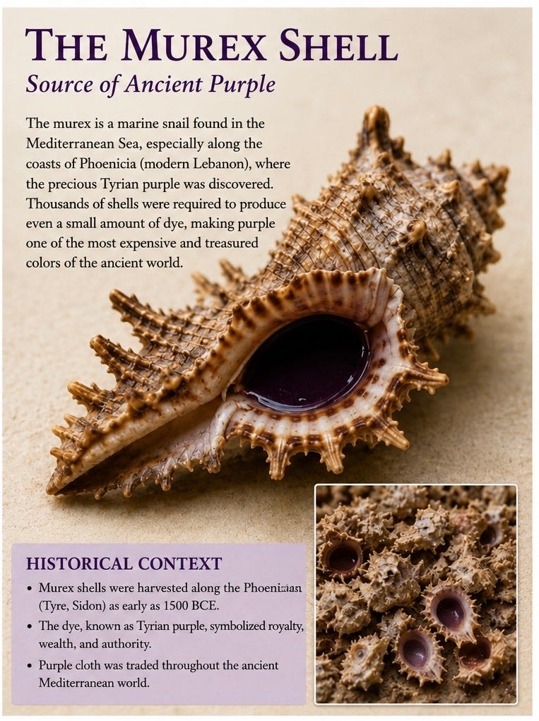
Purple dye was among the most prized substances in the ancient world.
The famous Tyrian purple was produced from Murex sea snails gathered along the Phoenician coast near Tyre and Sidon in the eastern Mediterranean.
Thousands of Murex shells were often required to produce a tiny amount of dye, making purple extraordinarily expensive and associated with royalty, nobility, and honor.
Phoenician merchants carried these dyes across maritime trade routes stretching from the Levant to Greece, Rome, North Africa, and beyond.
One of the most memorable figures connected to that world is Lydia of Thyatira.
The book of Acts introduces her this way:
“And a certain woman named Lydia, a seller of purple, of the city of Thyatira, which worshipped God, heard us: whose heart the Lord opened.” — Acts 16:14
Thyatira, located in Asia Minor (modern day Turkey) was known for textile production and dye guilds. Lydia’s trade likely connected her to these ancient commercial networks of costly dyed fabrics moving throughout the Roman world.
Before Scripture says anything about theology, it introduces her through hospitality, trade, beauty, craftsmanship, and color.
Lydia is the first recorded European convert to Christianity.
Scarlet and crimson carried their own sacred history.
Throughout the Hebrew Scriptures, scarlet thread and crimson dye appear in priestly garments, temple curtains, purification rituals, covenant imagery, and sacrifice. Some of these crimson dyes were associated with the tola worm (tolaʿath), whose vivid scarlet coloring became woven into the symbolic imagination of Israel.
The tabernacle itself was filled with these colors:
“Moreover thou shalt make the tabernacle with ten curtains of fine twined linen, and blue, and purple, and scarlet…” — Exodus 26:1
Priestly garments carried them as well:
“And they shall take gold, and blue, and purple, and scarlet, and fine linen.” — Exodus 28:5
Even cleansing rituals incorporated scarlet thread alongside cedar wood and hyssop.
Book of Psalms 22 later uses the word tola in one of Scripture’s most haunting passages of suffering:
“But I am a worm, and no man;a reproach of men, and despised of the people.” — Psalm 22:6
The tola worm released its scarlet stain only through crushing. Fixed to the wood, it poured itself out until its life was spent, leaving behind the crimson thread from which the ancient dye was gathered.
Early Christians saw in the tola a living witness to the suffering Messiah, the One lifted up upon the wood, bearing the crushing weight of sin and pouring out His life so that others might be covered in mercy rather than shame.
Perhaps this is part of why these colors still carry such weight beneath the microscope.
Scarlet once marked priesthood, sacrifice, cleansing, covenant, and redemption.
The microscope becomes more than a scientific instrument. It becomes a way of seeing hidden worlds through color.
And over time, the pathologist begins to realize that color itself carries memory.
Kings and temples.
Priests and sacrifice.
Blood and healing.
Life and death.
All still whispering quietly through purple, crimson, blue, and rose beneath the microscope.